• Lun - Ven : 9:00 - 18:00
  • 33 (0)5.57571005
  • info@synvec.fr

Posts by admin

Promising drug combination for advanced prostate cancer

A new drug combination may be effective in treating men with metastatic prostate cancer. Preliminary results of this new approach are encouraging and have led to an ongoing international study being conducted in 196 hospitals worldwide.

“We hope to find a well-tolerated and effective treatment to slow the progression of prostate cancer in men with advanced prostate cancer. The approach combines several drugs and attacks the cancer on several fronts,” said Dr. Fred Saad, researcher at the University of Montreal Hospital Research Centre (CRCHUM) and principal investigator of the study.

Antonio Paris, 59, is one of the patients participating in the CRCHUM. “Since I started the new treatment 14 months ago, my cancer first remitted and now is stable,” he said.

With early detection, prostate cancer is treatable. The vast majority of men recover. But it’s a different story for castration-resistant metastatic prostate cancer. In this most advanced form of the disease, the cancer progresses despite treatment options which include radiation therapy, hormone administration, and even removal of the prostate or testicles. Few options are available to halt the cancer that has spread outside the prostate and is resistant to hormone treatment. Since 2011, abiraterone acetate, sold under the name Zytiga®, has been administered orally in addition to standard hormone treatment. It blocks the production of testosterone, the male hormone, which acts as a fuel for cancerous tumours. “This treatment and other recent advances have extended the survival rates of men with the most advanced stages of prostate cancer while improving their quality of life. Life expectancy has increased from 18 months on average in 2004 to three years in 2015.

In a Phase 1 clinical trial, researchers tested the safety of a preliminary combination treatment of abiraterone acetate and another drug not yet approved for market, JNJ-56021927. Antonio Paris participated in this initial research phase. “I take 14 pills a day, and it’s going very well. I have the sweats and I’m tired a bit, but it doesn’t stop me from doing all kinds of activities and even renovations,” he said.

“Our trial conducted on forty patients indicate that this treatment is safe. The combined drugs are well tolerated and the treatment appears effective,” said Dr. Saad. Given these encouraging results, the U.S. Food and Drug Administration and Health Canada authorized the start of a Phase 3 clinical study. The trial is designed to compare the efficacy of abiraterone acetate (1000 mg) and a placebo versus treatment combining abiraterone acetate (1000 mg) and JNJ-56021927 (240 mg). In both groups, patients also receive small doses of prednisone to help the treatment and reduce the side effects of abiraterone acetate. Dr. Saad co-directs this randomized, double-blind trial involving 960 patients worldwide.

The findings of this international study will not be known for about another three years. The future treatment will therefore not be marketed for several years. But the researchers are enthusiastic. They hope this first combination treatment will successfully delay progression of the disease and prolong life. And above all, that it will improve the quality of life of patients with this devastating disease.

http://www.eurekalert.org/pub_releases/2015-11/uomh-pdc112415.php

Read more

Study reveals structure of tuberculosis enzyme, could offer drug target

A team of scientists, including several from the U.S. Department of Energy’s Argonne National Laboratory, have determined the structures of several important tuberculosis enzymes, which could lead to new drugs for the disease.

Tuberculosis, caused by Mycobacterium tuberculosis bacteria, has proved incredibly stubborn even in the age of powerful antibiotics, infecting about one third of all people worldwide. Treatment can take up to nine months. It has stealth properties that protect it from antibiotics; it can hide inside human cells, avoiding the body’s immune system while it waits for the opportune moment to multiply; and it’s very resourceful at acquiring resistance.

“What we have now may not work in a few years,” said Andrzej Joachimiak, an Argonne Distinguished Fellow, head of the Structural Biology Center, co-principal investigator at the Center for Structural Genomics of Infectious Diseases and a corresponding author on the new study.

In order to make new drugs, researchers need to search through the thousands of proteins in the bacterial world to find one that does something so important the bacterium can’t live without it—and then make a little block to match.

One such entry point might be IMPDH (inosine-5?-monophosphate dehydrogenase), which is part of a cellular process that controls the making of guanine nucleotides, one of the building blocks for DNA and RNA. It’s so essential that virtually all living organisms, including human and bacterial pathogens, have versions of it.

“What we discovered earlier this year is that the human and bacterial versions bind molecules differently,” Joachimiak said. “This is very important for finding a molecule to build a drug around—you don’t want to inhibit a human enzyme, just the pathogen one.”

Researchers have been interested in the mycobacterium IMPDH enzyme as a drug target for years, Joachimiak said, but haven’t been able to produce it well enough to study it.

The team observed that one part of the enzyme’s structure was particularly wobbly, so they engineered a version without it using resources at the Advanced Protein Characterization Facility and then then determined the structure employing synchrotron protein crystallography at the Advanced Photon Source, a DOE Office of Science User Facility (both at Argonne).

The modified version functions very similarly to the original, Joachimiak said, but is much easier to purify and crystallize for study.

Brandeis University professor Lizbeth Hedstrom and University of Minnesota professor Courtney Aldrich, two of the study’s other research collaborators, had identified several inhibitor molecules that bind to IMPDH, and thus might be a starting point for a drug—but they couldn’t be imaged while interacting with the enzyme. The new engineered enzyme allowed them to capture the structures of Hedstrom’s and Aldrich’s inhibitors in action, locked with IMPDH.

Helena Boshoff at the National Institute of Allergies and Infectious Diseases performed complementary studies showing that these inhibitors do in fact efficiently block mycobacterium growth.

The new structures were deposited into the Protein Data Bank for continued study

More information: Magdalena Makowska-Grzyska et al. Mycobacterium tuberculosis IMPDH in Complexes with Substrates, Products and Antitubercular Compounds, PLOS ONE (2015). DOI: 10.1371/journal.pone.0138976

Source link: http://phys.org/news/2015-11-reveals-tuberculosis-enzyme-drug.html#jCp

Read more

Maladie de PARKINSON : nilotinib, l’anticancéreux qui rebooste la cognition et la motricité

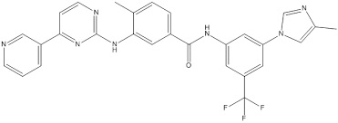
Ce petit essai clinique de l’Université de Georgetown dont les résultats viennent d’être présentés à Neuroscience 2015, la réunion annuelle de la Society for Neuroscience (Chicago), révèle le nilotinib (Tasigna®), un médicament indiqué dans le traitement de la leucémie myéloïde chronique (LMC) comme efficace pour améliorer la cognition et la motricité chez certains patients parkinsoniens. Alors que le médicament est déjà approuvé, ces données ouvrent un espoir à court terme pour les patients atteints et leur famille.

C’est un petit essai de phase I, d’une durée de 6 mois, de doses croissantes de nilotinib (150 à 300 mg par jour), mené sur 11 patients atteints de la maladie de Parkinson, montre sur 10 d’entre eux des améliorations cliniques significatives :

– l’efficacité sur la cognition, la motricité et l’amélioration d’autres fonctions (constipation) est observée chez la majorité des patients et de manière  » spectaculaire  » : ainsi, un patient, en fauteuil roulant s’est montré capable de remarcher. 3 autres patients ont retrouvé une fluidité de la parole. Le taux de chutes est également réduit.

Ces améliorations des symptômes sont corroborées par l’observation d’évolutions positives dans le liquide céphalo-rachidien (LCR) de biomarqueurs connus de la maladie de Parkinson (α-synucléine, bêta-amyloïde et tau, dopamine).

Le Dr Charbel Moussa, directeur du Laboratoire sur la démence et la maladie de Parkinson de Georgetown à l’origine de cette découverte des bénéfices du nilotinib dans le traitement des maladies neurodégénératives suggère que le traitement semble pouvoir inverser à un degré dépendant du stade de la maladie, le déclin cognitif et moteur chez les patients atteints de maladies neurodégénératives.

De premiers signaux très prometteurs: S’il reste certes à mener des études plus vastes, avec groupe témoin, avant confirmer ces effets positifs, cependant les premiers signaux sont prometteurs, à plus d’un titre :

         En particulier, le traitement induit une augmentation de la production de dopamine chez de nombreux patients, permettant, dans certains cas de réduire leurs doses de L-dopa ou d’autres médicaments dopaminergiques.

         De plus, cette utilisation du nilotinib, à des doses beaucoup plus faibles que pour le traitement de la LMC n’entraine pas d’effets secondaires sévères.

         le médicament pénètre dans la barrière hémato-encéphalique en quantités supérieures aux médicaments dopaminergiques.

         Enfin, ici à  » petite  » dose, le nilotinib induit une autophagie cellulaire suffisante pour  » nettoyer  » les cellules du cerveau sans entraîner leur mort (comme c’est en revanche le cas, pour les cellules cancéreuses, lorsqu’il est utilisé à plus fortes doses dans le traitement de la LMC).

Seul bémol, c’est un traitement cher, soit plus de 10.000 $/ mois pour 800 mg par jour (dose pour LMC). La dose utilisée dans cette étude était de 150 et 300 mg par jour.

Etude / Neuroscience 2015 17-Oct-2015 Communiqué Cancer drug improved cognition and motor skills in small Parkinson’s clinical trial (Visuel@ Georgetown University)

Read more

Malaria Protein Could Give Us A Broad Cancer Treatment

What do tumor cells, the placenta and malaria all have in common? A sugar, apparently, but not just any old sugar: one that might pave way for the development of broad treatment effective against a range of cancers.

This discovery, published in Cancer Cell, is the result of a collaboration between the University of British Columbia, the University of Copenhagen and the BC Cancer Agency, and finally offers an explanation for a curious observation that scientists long struggled to explain. Pregnant women, it turns out, are particularly susceptible to malaria. And it was the search for an explanation that ultimately led to this important find.

The researchers discovered that the malaria parasite, Plasmodium falciparum, engineers infected red blood cells to produce and present a malarial protein called VAR2CSA, which sticks to a type of sugar exclusively found in the placenta. Or so they thought. Tumor cells, it was revealed, also express a virtually identical sugar molecule. Both molecules are a type of chondroitin sulfate. That wasn’t a tremendous shock, though, because these two distinct tissues do share similarities: they both grow very rapidly and in an invasive manner.

“Scientists have spent decades trying to find biochemical similarities between placenta tissue and cancer, but we just didn’t have the technology to find it,” study coauthor and project leader Mads Daugaard said in a statement. “When my colleagues discovered how malaria uses VAR2CSA to embed itself in the placenta, we immediately saw its potential to deliver cancer drugs in a precise, controlled way to tumors.”

To examine this idea, the researchers generated VAR2CSA in the lab and attached one of two toxic molecules to it before testing out the novel compound on a bounty of both normal and tumor cell lines representing various different cancers. Encouragingly, the drug displayed specificity for the cancer cells only, attaching to them and delivering a deadly toxic blow after being absorbed. In fact, the compound was so potent that it successfully targeted and killed 95% of the cancer cell lines investigated, which included brain, blood, prostate and breast.

But sticking your finger in a tissue culture dish will quite easily kill cells, so the researchers took their drug one step further and tested it out on mice that were implanted with three different human cancers, all of which responded. In those with non-Hodgkin’s lymphoma, the tumors shrunk to around a quarter of the size of those in the untreated group, and two out of six treated mice with prostate cancer went into remission just one month after receiving the drug. Last but not least, five out of six mice with metastatic (tumors that have spread) breast cancer were cured.

“There is some irony that a disease as destructive as malaria might be exploited to treat another dreaded disease,” lead author Ali Salanti said in a statement.

Of course, we can’t get overexcited about animal trials, but the results are nonetheless encouraging. And two companies are already working towards developing a compound suitable for trials in humans, but that will take at least three to four years.


Source link : http://www.iflscience.com/health-and-medicine/malaria-protein-could-give-us-broad-cancer-treatment

Read more

Evidence for Person-to-Person Transmission of Alzheimer’s Pathology

Study raises question about whether proteins implicated in the disease are capable of spreading through medical procedures

Prions are the misshapen proteins that replicate by inducing normal proteins to misfold and aggregate in the brain, leading to rare diseases such as mad cow and kuru. In recent years, scientists have discovered that similar processes of protein misfolding are at work in many neurodegenerative disorders, including Alzheimer’s, Parkinson’s and Lou Gehrig’s disease. Now, a study in Nature reveals the first evidence for human-to-human transmission of the misfolded proteins that underlie the pathology of Alzheimer’s disease

The new findings draw upon earlier research conducted on a prion disease. Between 1958 and 1985, a number of individuals with short stature received shots of human growth hormone extracted from the pituitary glands of cadavers. The gland is a pea-sized structure that sits at the base of the brain. Some of these samples were contaminated with prions that caused certain patients to develop Creutzfeldt-Jakob disease (CJD), a rare and fatal brain disorder. Treatments ceased once these reports came to light, but by that time an estimated 30,000 people had already received the injections. As of 2012, researchers have identified 450 cases of CJD worldwide that are the result of these growth hormone injections and other medical procedures, including neurosurgery and transplants.

Misfolding of the amyloid-beta proteins is a hallmark of Alzheimer’s. Previous studies have shown that minute amounts of amyloid-beta injected into animals such as mice or monkeys act as seeds that initiate a chain reaction of protein misfolding that resembles the pathology of Alzheimer’s. However, until now, no studies have found evidence that this process occurs in humans.

To explore the question of human transmission, John Collinge, a neuroscientist at University College London and his colleagues, conducted an autopsy study of eight patients who died from CJD after treatment with cadaver-derived growth factor. To their surprise, they found that six of the brains had the amyloid-beta pathology found in Alzheimer’s patients, and four exhibited some degree of cerebral amyloid angiopathy, in which amyloid deposits build up on the walls of blood vessels in the brain.

The patients were between the ages of 36 and 51—typically too young to exhibit Alzheimer’s pathology—and none of the individuals bore genetic mutations associated with early onset of the disease. All evidence pointed toward one possibility: Like prions, amyloid-beta seeds were in the growth hormone injections and infected these individuals. Although none of the brains showed any other Alzheimer’s disease markers, such as buildup of another misfolded protein called tau, the researchers suggest that had the patients not died young, they would have developed the disease later in life.

The research may be a first step toward answering the question of whether human-to-human transmission of pathological proteins is possible. “This is an observational study,” Collinge says. “We’re simply describing what we see in these patients and we are trying to explain that.” This study alone, he says, does not suffice to prove that the Alzheimer’s disease process can be induced in one individual through contact with another’s brain tissue. In a follow-up study, the researchers are hoping to obtain archived batches of the cadaver-derived human growth hormone to look for the presence of telltale, small clusters of amyloidbeta.

By Diana Kwon

source link : http://www.scientificamerican.com/article/evidence-for-person-to-person-transmission-of-alzheimer-s-pathology/


Read more

Elastic drug delivery technology releases drugs when stretched

Researchers from North Carolina State University and the University of North Carolina at Chapel Hill have developed a drug delivery technology that consists of an elastic patch that can be applied to the skin and will release drugs whenever the patch is stretched.

For example, if applied to the elbow, the patch would release a drug when the elbow bends and stretches the patch.

“This could be used to release painkillers whenever a patient with arthritic knees goes for a walk, or to release antibacterial drugs gradually as people move around over the course of a day,” says Zhen Gu, co-senior author of a paper describing the work and an assistant professor in the joint biomedical engineering program at NC State and UNC-Chapel Hill.

The technology consists of an elastic film that is studded with biocompatible microcapsules. These microcapsules, in turn, are packed with nanoparticles that can be filled with drugs.

Here’s how it works: The microcapsules stick halfway out of the film, on the side of the film that touches a patient’s skin. The drugs leak slowly out of the nanoparticles and are stored in the microcapsules. When the elastic film is stretched, it also stretches the microcapsules – enlarging the surface area of the microcapsule and effectively squeezing some of the stored drug out onto the patient’s skin, where it can be absorbed.

“When the microcapsule is stretched from left to right, it is also compressed from bottom to top,” says Yong Zhu, co-senior author of the paper and an associate professor of mechanical and aerospace engineering at NC State. “That compression helps push the drug out of the microcapsule.”

After being stretched, the microcapsule is “re-charged” by the drugs that continue to leak out of the nanoparticles.

“This can be used to apply drugs directly to sites on the skin, such as applying anti-cancer medications to melanomas or applying growth factors and antibiotics for wound healing,” says Jin Di, co-lead author and a Ph.D student in Gu’s lab.

The researchers also incorporated microneedles into the system, applying them on top of the microcapsules. In this configuration, the drugs can be squeezed through the microneedles. The microneedles are small enough to be painless, but large enough to allow drugs to diffuse into the bloodstream through tiny capillaries underneath the skin. “This expands the range of drugs that can be applied using the technology,” says Shanshan Yao, co-lead author and a Ph.D student in Zhu’s lab.

“We’re now exploring how this tool can be used to apply drugs efficiently and effectively to burn patients, and we plan to look at how this could be used for pain relief as well,” Gu says.

“The materials are relatively inexpensive, and the manufacturing process is fairly straightforward, so we’re optimistic that this could be scaled up in a cost-effective way,” Zhu says.

The paper, “Stretch-Triggered Drug Delivery from Wearable Elastomers Containing Therapeutic Depots,” is published online in the journal ACS Nano. Co-authors include Yanqi Ye and Jicheng Yu of the joint biomedical engineering program, and Zheng Cui and Tushar Ghosh of NC State.

The work was supported by the American Diabetes Association, a pilot grant from UNC Chapel Hill’s NC TraCS Institute, and by the National Science Foundation through the ASSIST Engineering Research Center at NC State.

Source link : https://news.ncsu.edu/2015/08/technology-releases-drugs-when-stretched/

Read more

The FDA has just approved the first 3D-printed drug

We’re always hearing about how 3D printing will be capable of delivering major medical advancements  in the near future, but this time the big change is right on our doorstep: the US Food and Drug Administration (FDA) has just approved the first 3D-printed drug, potentially heralding a turning point in the way that modern medications are manufactured and taken.

Spritam, made by US-based pharmaceuticals company Aprecia, is an oral formulation of levetiracetam, which is used to control seizures in people who have epilepsy. This new 3D-printed incarnation of the drug is manufactured by a proprietary system developed by Aprecia called ZipDose, which stacks layer upon layer of powdered medication with liquid to create a new kind of highly disintegratable tablet

The technique ensures that the 3D printing isn’t just for marketing buzz – although that weren’t hurt Aprecia either. The idea is that the tiny tablet and rapid delivery of levetiracetam will make Spritam easier for people to take by encouraging quick, consistent and regular use.

While all ongoing medications should be taken as prescribed – which usually calls for following a regular dosage schedule – the consequences of forgetting to take or otherwise skipping epilepsy medication can be more severe than missing medications for other illnesses. Aprecia cites studies that have shown poor adherence to epilepsy medication can result in experiencing breakthrough seizures. In one case, 71 percent of patients surveyed admitted to having forgotten, missed, or skipped a dose of seizure medication, with almost half reporting a seizure resulting from the lapse.

The ZipDose system is designed to get around these sorts of issues by providing a quicker delivery form: a high dose of medication in a rapidly disintegrating tablet. It does this by depositing liquid droplets of an aqueous fluid on each thin layer of powdered medication. When the 3D-printed tablet comes into contact with a small amount of water, the bonds holding the pill together will rapidly break down, enabling a patient to consume as much as 1,000 mg of a medicated dose with just a single sip of water. It’s not a magic bullet ensuring people take their meds, but anything that makes pills easier to swallow can’t hurt patients’ prospects.

“By combining 3DP technology with a highly-prescribed epilepsy treatment, Spritam is designed to fill a need for patients who struggle with their current medication experience,” said Don Wetherhold, Aprecia’s CEO, in a statement. “This is the first in a line of central nervous system products Aprecia plans to introduce as part of our commitment to transform the way patients experience taking medication.”


Source link : http://www.sciencealert.com/the-fda-has-just-approved-the-first-3d-printed-drug

Read more

Biochemical sensor implanted at initial biopsy could allow doctors to better monitor, adjust cancer treatments

In the battle against cancer, which kills nearly 8 million people worldwide each year, doctors have in their arsenal many powerful weapons, including various forms of chemotherapy and radiation. What they lack, however, is good reconnaissance—a reliable way to obtain real-time data about how well a particular therapy is working for any given patient.

Magnetic resonance imaging and other scanning technologies can indicate the size of a tumor, while the most detailed information about how well a treatment is working comes from pathologists’ examinations of tissue taken in biopsies. Yet these methods offer only snapshots of , and the invasive nature of biopsies makes them a risky procedure that clinicians try to minimize.

Now, researchers at MIT’s Koch Institute for Integrative Cancer Research are closing that information gap by developing a tiny biochemical sensor that can be implanted in cancerous tissue during the initial biopsy. The sensor then wirelessly sends data about telltale biomarkers to an external “reader” device, allowing doctors to better monitor a patient’s progress and adjust dosages or switch therapies accordingly. Making cancer treatments more targeted and precise would boost their efficacy while reducing patients’ exposure to serious side effects.

“We wanted to make a device that would give us a chemical signal about what’s happening in the tumor,” says Michael Cima, the David H. Koch (1962) Professor in Engineering in the Department of Materials Science and Engineering and a Koch Institute investigator who oversaw the sensor’s development. “Rather than waiting months to see if the tumor is shrinking, you could get an early read to see if you’re moving in the right direction.”

Two MIT doctoral students in Cima’s lab worked with him on the sensor project: Vincent Liu, now a postdoc at MIT, and Christophoros Vassiliou, now a postdoc at the University of California at Berkeley. Their research is featured in a paper in the journal Lab on a Chip that has been published online.

Measurements without MRI

The developed by Cima’s team provide real-time, on-demand data concerning two biomarkers linked to a tumor’s response to treatment: pH and dissolved oxygen.

As Cima explains, when is under assault from chemotherapy agents, it becomes more acidic. “Many times, you can see the response chemically before you see a tumor actually shrink,” Cima says. In fact, some therapies will trigger an immune system reaction, and the inflammation will make the tumor appear to be growing, even while the therapy is effective.

Source : http://phys.org/news/2015-08-biochemical-sensor-implanted-biopsy-doctors.html#jCp

Read more

‘Single-molecule Tetris’ allows scientists to observe DNA at the nanoscale

Physicists are using a technique reminiscent of a classic video game to observe DNA on the nanoscale. They call it “single-molecule Tetris.” The approach consists of a device filled with tiny channels and cavities that DNA molecules can move in and out of, resulting in some of the familiar Tetris shapes, like the “L,” the square, and the zigzag (illustrated above: a DNA molecule, in red, occupies four cavities in a zigzag). As the chainlike molecules bend or jump into different shapes, researchers use that information to measure two very specific characteristics of DNA molecules—the width and the confined free energy, or entropy of the molecule. Here, entropy relates to how many ways the energy of the molecule can be arranged. Physically, it correlates to the different shapes the molecule can take while maintaining the same distance between its two ends. Scientists collected data, published this month in Macromolecules, on these parameters to show the precise conditions under which DNA can be trapped in the cavities. It’s a feat that’s extremely difficult to do on such a minute scale, but thanks to single-molecule Tetris, these physicists now have results that could help biologists improve genome sequencing and tease out valuable genetic information from these tiny, confined bits of DNA.

Source link : http://news.sciencemag.org/chemistry/2015/07/single-molecule-tetris-allows-scientists-observe-dna-nanoscale

Read more

Studies confirm regorafenib benefit in pre-treated metastatic colorectal cancer

The Phase IIIb CONSIGN study has confirmed the benefit of regorafenib in patients with previously treated metastatic colorectal cancer (mCRC).

Researchers announced the results at the European Society for Medical Oncology (ESMO) World Congress on Gastrointestinal Cancer 2015 in Barcelona

The safety profile and progression free survival were similar to Phase III trial results.

CONSIGN was a prospective, observational study that was initiated to allow patients with mCRC access to regorafenib before marketing authorisation and to assess safety, which was the primary endpoint. The randomised Phase III CORRECT trial previously showed that regorafenib significantly improves survival in patients with pre-treated mCRC and led to regulatory approval.

“We began CONSIGN at the suggestion of the authorities and to fulfil the wishes of patients and doctors for a larger expanded access,” said lead study author Prof Eric Van Cutsem from University Hospitals Leuven, Belgium. “Today we report on safety and progression-free survival in a large cohort of patients that more closely resembles daily clinical practice than the pivotal registration trial.”

CONSIGN included more than 2,800 patients at 188 sites in 25 countries who received regorafenib for a median of 2.5 months. Grade >3 adverse events occurred in 80% of patients. The estimated progression-free survival was 2.7 months and was similar across KRAS wild type and mutant subgroups.

Need to establish guidelines on the management of adverse events to make regorafenib more tolerable for patients

Van Cutsem said, “This study in a real world population of patients with pre-treated mCRC shows a similar safety profile and progression-free survival with regorafenib as shown in the randomised CORRECT trial. The findings add to our knowledge of how to select patients and how to manage toxicities. We need to establish clear guidelines on the management of adverse events to make taking the drug more tolerable for patients.”

Commenting on the data, Dr Dirk Arnold, ESMO spokesperson, director of the Department of Medical Oncology, Klinik für Tumorbiologie in Freiburg, Germany, said, “CONSIGN confirms the efficacy and safety data of the randomised Phase III CORRECT and CONCUR trials. The merit of CONSIGN is that it translates Phase III data into the clinical routine since patients had similar characteristics and pre-treatment to what we see in daily practice.”

The adverse events reported in CONSIGN were within the scope of expectation and comparable to the CORRECT trial, added Arnold. “There were no surprising findings in terms of toxicity,” he said. “All of the adverse events were quite class specific and also likely manageable.”

He added, “CONSIGN depicts what we would expect from an observational trial in this setting. It shows that we have further treatment options for mCRC patients pre-treated with chemotherapy, and that this comes at the cost of a specific, but manageable toxicity profile.”

Regarding the next step in this research area, Arnold said, “Biomarkers have been extensively investigated in the randomised trials but until now nothing has been found that would allow prediction of the benefit of regorafenib for a specific group of patients. I would suggest having a further look at the data in the observational CONSIGN study to see if there are clinical characteristics that identify patients who could benefit more or less from this treatment.”

Source : http://www.europeanpharmaceuticalreview.com/33044/news/industry-news/studies-confirm-regorafenib-benefit-in-pre-treated-metastatic-colorectal-cancer/


Read more